Blog
Advances in Digital Pathology
June 2, 2026

Advances in technology are increasingly automating and digitizing healthcare processes and laboratory information systems. These advancements have helped speed clinical diagnosis and treatment, improving patient outcomes across the healthcare continuum.
One such emerging technology is digital imaging, which has transformed digital pathology by enabling high-throughput scanning of patient samples.
The traditional way of preparing a tissue sample for investigation and LIS pathology lab workflow involves a histology tech segmenting the sample into thin slices and mounting them onto glass slides. These are then processed to improve final image quality before being sent to the pathologist, who examines the samples under a microscope to determine or confirm a diagnosis.
While these steps are still largely followed, digitization has begun to transform LIS systems and anatomic pathology software modules for optimized pathology lab management by converting the glass slide prepared for microscopy into a more flexible digital image within an exciting new digital pathology workflow.
Discover More: The Role of Anatomic Pathology LIS Software in Optimizing Laboratory Workflow Management
Defining Pathology Lab Management with the Benefits of Digital Pathology Solutions
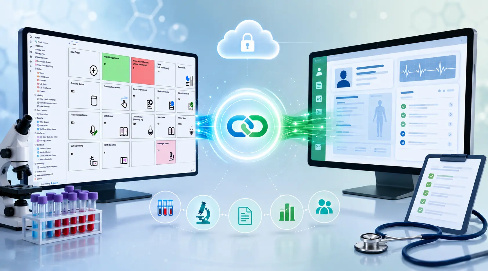
Digital pathology is made possible by digitizing prepared slides and enabling an integrated digital pathology and LIS lab workflow, replacing glass slides traditionally viewed under a microscope with digital images that can be scanned and viewed on a monitor.
A digital pathology scanner replaces the microscope by capturing whole-slide images and transmitting them directly to the pathologist's screen via the lab information system.
Discover More: Pathology Lab Reporting Software and Pathology Lab Management: Enhancing Laboratory Efficiency
Key Benefits for Progressive Pathology Groups
The pathologist can share digital pathology images instantly with the medical team, eliminating the need to ship slides by courier between sites. It speeds up turnaround, streamlines operations, fosters better collaboration, and ultimately improves patient care.
Discover More: Introducing LigoLab’s Grossing Workstation Window

Digital Pathology Workflow with Image Scanners
How Digital Pathology Scanners Work
A histology or digital pathology scanner can process up to 1,000 glass slides at one time, capturing high-resolution digital images and displaying them on a connected workstation. To maintain image quality, scanners should be positioned on stable laboratory countertops away from vibrations generated by nearby equipment such as centrifuges or stirrers. Each scanner integrates with a digital pathology viewing environment through connected monitors and linked image management systems (IMS) and laboratory information system (LIS) software applications.
Lab managers can choose digital pathology scanners based on their routine application in the lab. For example, if a pathologist needs images with high magnification, such as 40X, the appropriate diagnostic lab software and scanner can be selected to achieve that level of detail.
Discover More: Executive War College 2026 Recap - Advancing Laboratory Leadership Through Innovation, Collaboration, and Digital Transformation
Speed, Volume, and Customization
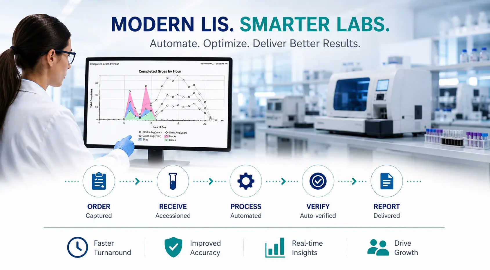
Today's digital pathology scanners can capture and process images within a minute, adjust them to multiple magnifications, and handle large volumes of slides. Image acquisition through scanning is accelerated with today's advanced digital pathology solutions, improving turnaround time for clinical diagnosis within laboratory information systems and enhancing overall clinical lab workflow and pathology lab management.
The best digital pathology scanners are customizable to a lab's needs. Two common customization options include:
Complete Automation: A fully automated process of capturing whole slide images, from slide preparation and management to diagnosis and reporting, with minimal or no human intervention.
Semi-Automation: A semi-automated process that integrates manual and automated steps, combining human expertise with digital tools for preparation, management, diagnosis, and reporting.
Scanners should also be compatible with modern pathology lab software applications, such as advanced laboratory information systems that automatically acquire, label, and store digital images.
Discover More: Comparing LigoLab Informatics Platform with Legacy Laboratory Information System Software
DP Workflow with Pathology Image Analysis Software
AI-Driven Analysis and Pattern Recognition
Through pathology image analysis software, labs can leverage artificial intelligence (AI) analytical tools and algorithms to streamline a pathologist's lab workflow and decrease the chance of human errors that may arise during sample processing.
Analysis of acquired images is semi-automated using modern digital pathology software, enabling a pathologist to investigate a slide by directly annotating the image with measurements recorded in an advanced lab information system. Image acquisition management and pattern analysis on digitized images enable the interpretation of pathology data for clinical diagnosis.
Historical Data Comparison and Diagnostic Confidence
Pathologists can evaluate and compare their diagnoses with historical data stored and processed within the same pathology image analysis software or in an integrated lab information system. Digital pathology solutions thus support reproducible interpretations, empirical measurements, and greater confidence in pathologists' findings.
Discover More: Pathology Software and Laboratory Information Technology

The Many Benefits of Digital Pathology Solutions
Anatomic pathology labs are adopting laboratory software systems that support advanced digital pathology solutions for numerous reasons:
- Instant image sharing simplifies collaboration among a team of pathologists, accelerating diagnosis times and enabling second opinions through online transmission.
- Secure cloud storage enables digital pathology images to be archived instantly and permanently, improving accessibility for peer consultations while keeping patient information safe.
- Embedded analysis tools measure specific tissue biomarkers that indicate disease, with AI guiding the review and improving diagnostic accuracy.
- Barcode labeling matches slides to patient information, significantly reducing the risk of misidentification.
- Faster diagnoses enable patients to receive results more quickly, even as case volumes increase.
- Pathologist development benefits from richer interaction with collected sample data.
- Flexible work schedules become possible as digital pathology provides a solid foundation for workflow automation.
- LIS lab integration enables image acquisition software and digital pathology workflows to connect directly with laboratory information system software deployed within the lab.
- Reduced storage costs result from archiving digital images rather than maintaining physical slides or frozen sections.
- Eliminating sample degradation ensures that digital images remain pristine over time, unlike physical specimens.
Case Study: Summit Pathology - Achieving Laboratory Profitability Amidst Operational Pressures

The Integration of Digital Pathology Solutions and Advanced LIS Systems
Today’s digital pathology solutions have advanced rapidly, and one key area of progress has been the seamless integration of digital pathology applications with advanced laboratory information systems. Labs that operate with modern pathology LIS systems are at a significant advantage when adding digital pathology to their workflow.
Key Advantages of Digital Pathology and LIS Software Integration
Efficient Laboratory Workflow Management: Digital pathology integration streamlines pathology lab management by enabling seamless data and image transfer between integrated laboratory software systems, reducing manual steps, minimizing errors, and accelerating diagnostic processing.
Enhanced Accessibility: Digital pathology enables remote access to images and data, facilitating strong collaboration among pathologists regardless of location and adding a layer of consultation that was previously impossible to achieve.
Improved Accuracy: Advanced image analysis tools assist pathologists in diagnosis, while integration with the best laboratory information system software ensures that all relevant patient data is readily available during analysis, leading to more accurate and comprehensive diagnostic reports.
Centralized Data Management: The best laboratory information system software also enables centralized storage and management of anatomic pathology LIS data within the system database, improving data integrity, security, and accessibility while simplifying retrieval for audits and quality assurance.
Cost Savings: Long-term benefits include reduced storage costs for physical slides, improved resource utilization, and potential savings from faster turnaround times and more accurate diagnoses.
Scalability: Digital pathology solutions integrated with modern medical LIS systems can easily scale to accommodate growing volumes of cases and data, enabling laboratories to adapt to increasing workloads without compromising efficiency or quality.
Compliance: Digital pathology integration with modern LIS medical solutions helps laboratories comply with regulatory requirements by ensuring proper documentation, traceability, and data security, simplifying the accreditation process and reducing compliance risk.
Patient-Centric Care: By providing quicker access to pathology results, digital pathology integration with today’s leading anatomic pathology LIS systems supports faster treatment decisions, improved patient outcomes, and greater patient satisfaction.
Discover More: LigoLab’s Anatomic Pathology LIS Lab Solutions
The Future of Digital Pathology Solutions
The U.S. healthcare system is under pressure from the massive amount of data produced during diagnostics. To keep pace, labs need advanced digital pathology solutions that streamline workflow and handle growing demands. Researchers and clinicians increasingly apply machine learning and big data to diagnostic pathology, enabling more precise and efficient analysis.
Emerging Capabilities
Today’s digital pathology systems can perform complex tasks such as multiplex analysis, measuring multiple markers within the same tissue sample, and enabling accurate comparison of signal intensities between markers. This level of analysis is extremely difficult to perform manually under a microscope due to stain diffusion.
These solutions also open integration with other diagnostic methods. For instance, biomarker expression identified through digital pathology can be compared with protein expression data from mass spectrometry, creating a more comprehensive diagnostic picture.
A Transforming Field
With improved image management systems, modern integration with laboratory information systems, and evolving data-sharing policies, the potential for digital pathology continues to expand. Cloud storage, virtual microscopy, and AI-driven tools are transforming how pathologists work, replacing older manual methods with faster, more collaborative, and more accurate diagnostics.
On-Demand Webinar: Learn How Modern Anatomic Pathology LIS Software Can Transform Your Lab’s Operations
Ready to Integrate Digital Pathology and Advanced Laboratory Information System Software Into Your Lab Workflow?
Take Action: Speak with a LigoLab Product Specialist Today!
Frequently Asked Questions About Today’s Digital Pathology Solutions and Integration with Advanced Laboratory Information Systems
What is digital pathology, and how does it differ from traditional microscopy?
Digital pathology replaces the traditional microscope workflow by scanning prepared glass slides into high-definition digital images, known as whole slide images, that users can view, annotate, share, and store electronically. Unlike traditional microscopy, digital pathology enables remote consultation, AI-assisted analysis, instant image sharing, and secure cloud archiving.
How does a digital pathology scanner work?
A digital pathology scanner captures high-resolution whole-slide images from prepared glass slides and transmits them to a connected computer screen via laboratory information system software. Modern scanners can process images within a minute, handle up to 1,000 slides at a time, and adjust images to multiple magnification levels, supporting both fully automated and semi-automated workflows.
What is the difference between fully automated and semi-automated digital pathology workflows?
Fully automated workflows handle the entire process, from slide preparation and image capture to diagnosis and reporting, with minimal human intervention. Semi-automated workflows combine automated tools with manual steps, leveraging human expertise at key stages while still benefiting from digital efficiency and accuracy improvements.
How does AI-driven image analysis improve digital pathology workflows?
AI-based analytical tools and algorithms can automatically analyze acquired digital images, detect patterns, measure tissue biomarkers, and flag areas of interest. It reduces human error during sample processing, supports reproducible diagnostic interpretations, and enables pathologists to compare results with historical data stored in the lab information system, thereby increasing diagnostic confidence.
What are the key benefits of integrating digital pathology with an advanced LIS system?
Key benefits include streamlined laboratory workflow management through seamless data and image transfer, enhanced remote accessibility for pathologist collaboration, improved diagnostic accuracy through integrated image analysis tools, centralized data management with improved security and audit readiness, reduced storage costs, scalability for growing case volumes, regulatory compliance support, and faster patient results leading to better clinical outcomes.
How does digital pathology reduce storage costs compared to traditional slide management?
Physical glass slides require dedicated storage space, degrade over time, and pose risks of fire, theft, or damage. Digital pathology replaces physical archives with secure cloud storage, eliminating the cost and risk of maintaining large physical slide collections while keeping images permanently accessible and unaffected by physical deterioration.
What emerging technologies are shaping the future of digital pathology?
Machine learning, big data analytics, and AI-driven diagnostic tools are enabling more precise and efficient pathology analysis. Multiplex analysis enables digital pathology systems to measure multiple markers in a single tissue sample, a task that is extremely difficult to perform manually. Integration with other diagnostic methods, such as mass spectrometry, is also expanding the comprehensiveness of pathology diagnoses.